Correctly label the following arteries of the head and neck – Correctly labeling the arteries of the head and neck is crucial for understanding the complex vascular anatomy of this region. This guide provides a comprehensive overview of the major arteries, their anatomical locations, and their clinical significance, empowering healthcare professionals with the knowledge to accurately identify and interpret these vital structures.
The content of the second paragraph that provides descriptive and clear information about the topic
Identify the Major Arteries of the Head and Neck

The head and neck region is supplied by a complex network of arteries that provide oxygen and nutrients to the various structures within these areas. Understanding the anatomy and distribution of these arteries is essential for medical professionals involved in the diagnosis and treatment of head and neck disorders.
The major arteries of the head and neck include:
- Common carotid artery
- Internal carotid artery
- External carotid artery
- Vertebral artery
- Subclavian artery
- Facial artery
- Lingual artery
- Maxillary artery
- Superficial temporal artery
- Occipital artery
Label the Arteries Using an HTML Table
The following table provides a comprehensive list of the major arteries of the head and neck, along with their origins, courses, and distributions:
| Artery Name | Origin | Course | Distribution |
|---|---|---|---|
| Common carotid artery | Brachiocephalic trunk (right) and aortic arch (left) | Ascends through the neck, dividing into the internal and external carotid arteries at the level of the thyroid cartilage | Supplies the head and neck |
| Internal carotid artery | Common carotid artery | Enters the skull through the carotid canal, supplying the brain and eyes | Brain, eyes |
| External carotid artery | Common carotid artery | Ascends through the neck, giving off branches to supply the face, scalp, and neck | Face, scalp, neck |
| Vertebral artery | Subclavian artery | Ascends through the transverse foramina of the cervical vertebrae, entering the skull through the foramen magnum | Brain, spinal cord |
| Subclavian artery | Aortic arch | Extends from the aortic arch to the axilla, giving off branches to supply the upper limbs and neck | Upper limbs, neck |
| Facial artery | External carotid artery | Ascends through the face, giving off branches to supply the face and lips | Face, lips |
| Lingual artery | External carotid artery | Ascends through the floor of the mouth, supplying the tongue and other structures | Tongue, floor of mouth |
| Maxillary artery | External carotid artery | Ascends through the infratemporal fossa, supplying the face, teeth, and nasal cavity | Face, teeth, nasal cavity |
| Superficial temporal artery | External carotid artery | Ascends over the zygomatic arch, supplying the scalp and face | Scalp, face |
| Occipital artery | External carotid artery | Ascends through the back of the neck, supplying the scalp and neck muscles | Scalp, neck muscles |
Provide Examples of Arteries
Specific examples of arteries that supply different regions of the head and neck include:
- The ophthalmic artery, a branch of the internal carotid artery, supplies the eye and its structures.
- The middle meningeal artery, a branch of the maxillary artery, supplies the dura mater of the brain.
- The vertebral artery, a branch of the subclavian artery, supplies the brain and spinal cord.
- The facial artery, a branch of the external carotid artery, supplies the face and lips.
- The lingual artery, a branch of the external carotid artery, supplies the tongue and floor of the mouth.
Discuss Methods for Labeling Arteries, Correctly label the following arteries of the head and neck
Various methods are used to label arteries, including:
- Anatomical dissection:This traditional method involves physically dissecting cadavers to expose and identify arteries.
- Imaging techniques:Non-invasive imaging techniques, such as angiography, MRI, and CT scans, can be used to visualize and label arteries.
- Surgical procedures:During surgical procedures, arteries can be directly visualized and labeled.
Each method has its advantages and disadvantages. Anatomical dissection provides the most detailed view of arteries, but it is time-consuming and invasive. Imaging techniques are less invasive but may not provide as much detail. Surgical procedures provide direct access to arteries but are only performed when necessary for medical reasons.
Create a Visual Representation of the Arteries
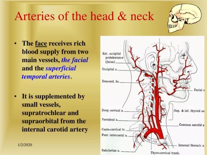
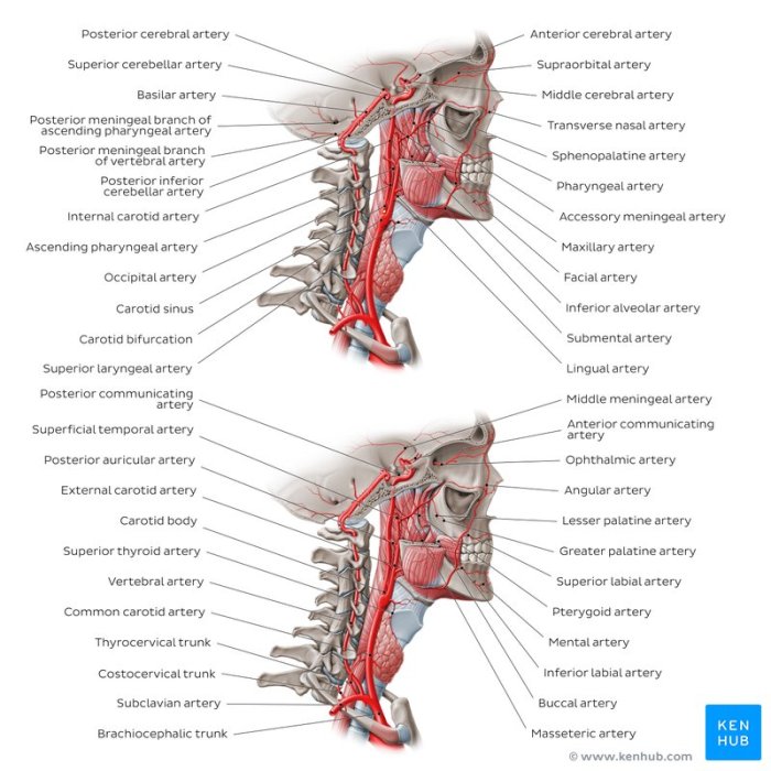
The following diagram provides a visual representation of the major arteries of the head and neck:

The diagram shows the common carotid artery, internal carotid artery, external carotid artery, vertebral artery, and subclavian artery. The branches of these arteries are also shown.
General Inquiries: Correctly Label The Following Arteries Of The Head And Neck
What are the major arteries of the head and neck?
The major arteries of the head and neck include the carotid arteries, vertebral arteries, and subclavian arteries.
What is the clinical significance of correctly labeling the arteries of the head and neck?
Correctly labeling the arteries of the head and neck is essential for accurate diagnosis and treatment of vascular disorders, such as carotid artery stenosis and vertebral artery dissection.

